")


Biochemistry and Molecular Biology
Faculty Sponsor: Jean Kennedy
School: Quinsigamond Community College
Research Area: Biochemistry and Molecular Biology
Location: Poster Session 1, 10:30 AM - 11:15 AM: Campus Center Auditorium [A5]
The use of brain scans and genetic evaluation shows strong potential to increase the precision of mental health diagnosis and treatment. Artificial intelligence, high-throughput sequencing, gene writing, and gene editing have enabled the introduction of genomic treatment and precision medicine. Five underlying genomic factors associated with psychiatric illness have been identified. These genetic signatures are unique to those that struggle with mental disorders and indicate biological similarities among distinct psychiatric disorders. Identifying patterns of brain connectivity using high-resolution imaging and advanced computational modeling with collaborative datasets can help determine the effectiveness of different types of antidepressant medications for patients with major depressive disorder. Diagnosis and treatment may also be improved through the analysis of predictive brain imaging biomarkers. Artificial intelligence tools are being utilized to develop digital biomarkers that support psychiatric research. The approach to and process of mental health diagnosis and treatment will be altered by new technologies. Advancements in neuroscientific understanding are likely to shape the future of psychiatry, offering the potential for more precise and personalized diagnostic approaches and treatments for mental illness.
RELATED ABSTRACTS
- Daydreaming Frequency as a Potential Modulator of TMR Effectiveness, Temple, Chloe, UMass Amherst, Poster Session 1, 10:30 AM - 11:15 AM, 163, C19
- LRP1-Mediated Tau Endocytosis in Alzheimer’s Disease, Trivedi, Samay Darshin, UMass Amherst, Poster Session 5, 3:15 PM - 4:00 PM, Auditorium, A53
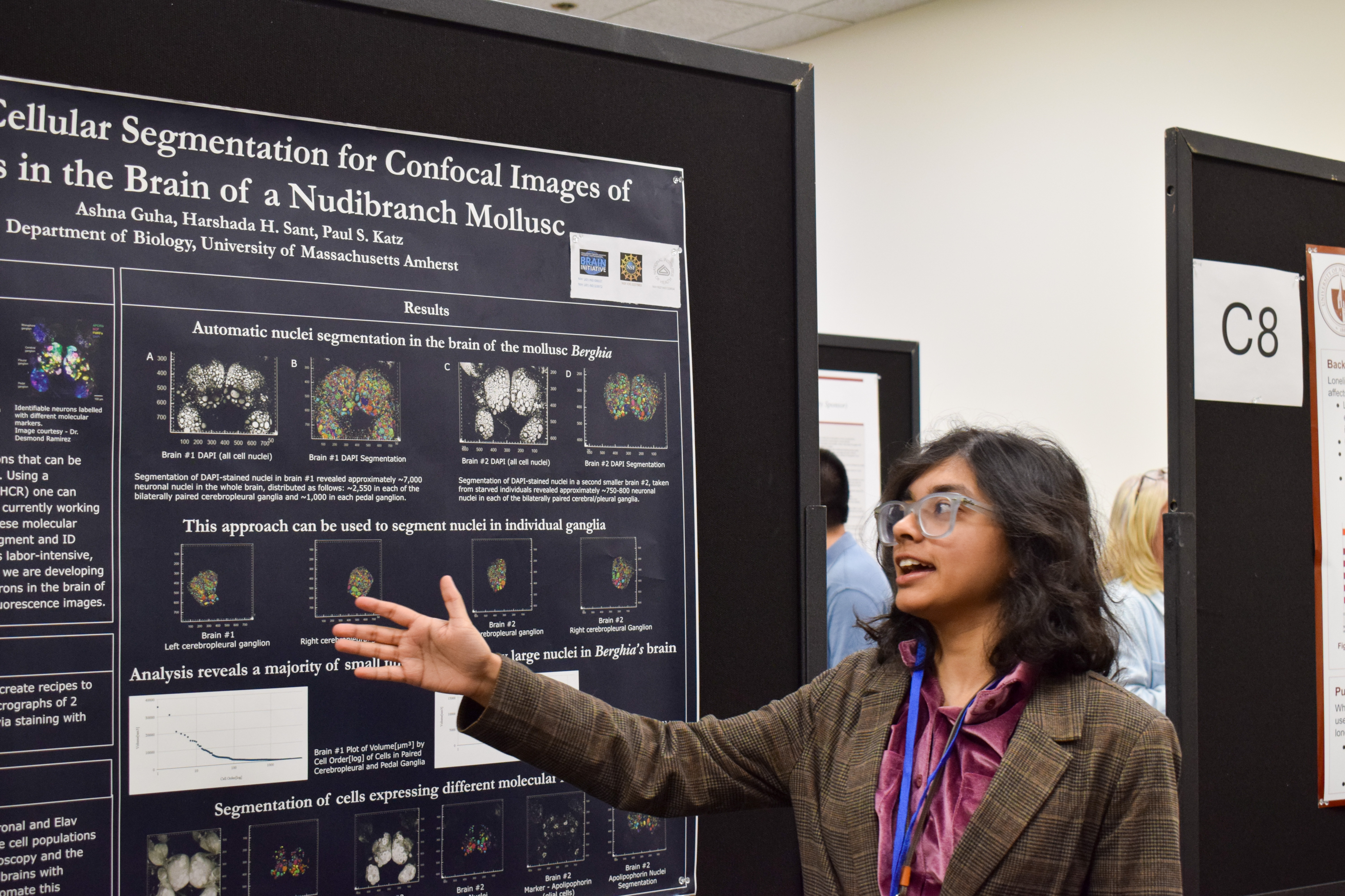
- How Does a Sea Slug Smell? A Molecular Approach to Investigating Olfaction in a Nudibranch, Thattacherry, Tess, UMass Amherst, Poster Session 5, 3:15 PM - 4:00 PM, Concourse, B2
- Environmental DNA (eDNA) as a Tool for Rare Freshwater Mussel Detection and Restoration Planning, Stratton, Mickala Diane, UMass Amherst, Poster Session 5, 3:15 PM - 4:00 PM, Auditorium, A40
- Synteny and Genetic Conservation of the Nicotinamide Amidase Gene in the Drosophilawillistoni and Drosophila bipectinata Species as Compared to Drosophila melanogaster, Lacina, Colbie , Quinsigamond Community College, Poster Session 1, 10:30 AM - 11:15 AM, Auditorium, A10
Faculty Sponsor: Vincent Rotello
School: UMass Amherst
Research Area: Biochemistry and Molecular Biology
Location: Poster Session 1, 10:30 AM - 11:15 AM: Concourse [B13]
Triple negative breast cancer (TNBC) is an aggressive breast cancer subtype characterized by the absence of estrogen receptor, progesterone receptor, and human epidermal growth factor receptor 2 (HER2) expression. Because TNBC lacks these therapeutic targets, current treatment relies primarily on non-specific chemotherapies, which cause substantial systemic toxicity and fail to prevent high rates of recurrence and metastasis. Enzyme prodrug therapy has emerged as a promising treatment for cancer, enabling localized activation of cytotoxic agents within tumors to minimize off-target toxicity. While this approach is promising, current delivery strategies like antibody-enzyme conjugates and vectors coding for the prodrug-activating enzyme struggle with inefficient delivery, immunogenicity, and uneven vector expression.
It has been demonstrated that Poly(oxanorborneneimide) polymers (PONI) featuring cationic guanidinium (Guan) moieties are capable of high efficiency (≥90%) direct cytosolic enzyme delivery while retaining enzymatic activity. Furthermore, the addition of a sulfobetaine zwitterionic block (Zwit) significantly improved tumor accumulation and slowed clearance by the reticuloendothelial system. Using the diblock copolymer PONI-Guan-Zwit, E. coli recombinant oxygen insensitive nitroreductase NfsB will be delivered to catalyze the activation of the DNA crosslinking prodrug 5-(aziridin-1-yl)-2,4-dinitrobenzamide (CB1954). Through in vitro delivery and functional characterization, polymer-directed enzyme prodrug therapy will be evaluated as a targeted and potentially less toxic therapeutic strategy for triple negative breast cancer.
RELATED ABSTRACTS
- Development of an Efficient One-Pot Synthesis of Phenoxathiin-10,10-Dioxide Derivatives for Prostate Cancer Research, Zou, Alice Bian, UMass Amherst, Poster Session 5, 3:15 PM - 4:00 PM, 165, D12
- Design and the Development of Protein Kinase C Inhibitors as Anticancer Agents, Furlong, Sean James, Worcester State University, Poster Session 2, 11:30 AM - 12:15 PM, Concourse, B16
- Lifestyle Strategies for a Healthy Autonomic Nervous System, Miller, Jalyn Allison, Springfield Technical Community College, Poster Session 2, 11:30 AM - 12:15 PM, Auditorium, A28
- Improving Undergraduate Student Genetics Literacy Through a Case Study Addressing Central Dogma Misconceptions, Letowska, Michelle, UMass Amherst, Poster Session 6, 4:15 PM - 5:00 PM, Auditorium, A50
- Evaluating Therapeutic Drug Candidates To Restore Dysferlin Function in Dysferlinopathy, Kwakye, Kaissy Obeng, Fitchburg State University, Poster Session 2, 11:30 AM - 12:15 PM, Auditorium, A68
Faculty Sponsor: Vincent Rotello
School: UMass Amherst
Research Area: Biochemistry and Molecular Biology
Location: Poster Session 1, 10:30 AM - 11:15 AM: Concourse [B14]
Engineered antimicrobial polymers provide a promising tool for fighting biofilm infections. The emergence of antimicrobial-resistant (AMR) infections caused by multidrug-resistant (MDR) bacteria poses a growing threat to global health. When left untreated, these pathogens can become more resistant by forming biofilms that can exacerbate chronic wound infections that can lead to sepsis. Biofilms form when different strains of bacterial cells aggregate and produce an extracellular matrix. The biofilm extracellular matrix is composed of extracellular polymeric substances that form intermolecular interactions, effectively protecting biofilm bacteria by further reducing the penetration and activity of antimicrobial agents and conventional antibiotics. Thus, the development of novel antimicrobial agents for the treatment of biofilm infections is urgently needed. This project presents highly effective engineered antimicrobial polymers. Engineered antimicrobial polymers offer alternative approaches to conventional antimicrobial agents and antibiotics, due to their tunable hydrophobic moieties. With careful engineering of hydrophobic and cationic domains, antimicrobial polymers effectively eradicate both planktonic bacteria and its biofilms counterpart. We explored a library of antimicrobial polymers with different degrees of hydrophobicity. Our amphiphilic polymers showing promising activity against planktonic MDR bacteria and excellent biofilm eradication. Importantly, engineered antimicrobial polymers facilitate membrane disruption mechanisms that can effectively combat bacterial resistance. Overall, engineered antimicrobial polymers provide an effective alternative to conventional antimicrobial agents for treating bacterial infections.
Faculty Sponsor: Vincent Rotello
School: UMass Amherst
Research Area: Biochemistry and Molecular Biology
Location: Poster Session 1, 10:30 AM - 11:15 AM: Concourse [B15]
Faculty Sponsor: Vincent Rotello
School: UMass Amherst
Research Area: Biochemistry and Molecular Biology
Location: Poster Session 1, 10:30 AM - 11:15 AM: Concourse [B16]
Biofilms are protected by a dense extracellular polymeric substance (EPS) matrix that shields bacteria from antibiotics, environmental stress, and host immunity. On medical devices such as catheters, biofilms become especially problematic, causing persistent infections that often require prolonged therapy or device replacement and accelerating antibiotic resistance. Essential oils have antimicrobial activity, but limited EPS penetration due to their poor solubility. This honors thesis evaluated gelatin-stabilized essential-oil nanoemulsions containing carvacrol (C-GNE), eugenol (E-GNE), or geraniol (G-GNE) against MRSA biofilms on catheters, surgical gauze, and gloves. In a 15-min inhibition assay, C-GNE yielded no detectable CFU/mL, while E-GNE and G-GNE produced ~5–6 log10 CFU/mL. Against 1-day-old biofilms treated for 3 h, C-GNE reduced bacterial counts by ~3–4 log10, whereas E-GNE and G-GNE achieved ~2–3 log10 reductions. Overall, C-GNE appears most promising as a natural, polymer-encapsulated antibiofilm approach for medical-device–associated infections. As the project continues, our central hypothesis is that these nanoemulsions inhibit and eradicate MRSA biofilms in a dose-dependent manner. Future work will focus on translating this system into a sanitizing spray or biodegradable disinfectants for the elimination of medical device-associated infections, where applications of conventional chemical disinfectants are limited or unsuitable.
Faculty Sponsor: Joslyn Mills
School: Bridgewater State University
Research Area: Biochemistry and Molecular Biology
Location: Poster Session 2, 11:30 AM - 12:15 PM: Room 163 [C28]
The C. elegans protein Lipid-binding protein 3 (LBP-3) is a lipid chaperone which transports polyunsaturated fatty acids (PUFAs) freed by lipolysis of intestinal fat deposits. This lipid signaling pathway involves the interaction of LBP-3 with the nuclear hormone receptor, NHR-49, a process which promotes transcription of life-extending neuropeptides, promoting longevity. A related protein in the nuclear hormone receptor family that could also potentially interact with LBP-3, NHR-86, has been identified as a transcription factor which upregulates immune-related gene expression, drawing question to a potential relationship between lbp-3 and the nematode’s immune response. It is predicted that reduced expression of lbp-3 causes lower levels of NHR-86 activation, impairing the organism’s innate immunity.
To test this hypothesis, physiological, quantitative, and computational experiments were conducted. Physiologically, the effect of reduced lbp-3 expression on the immune system of C. elegans was examined through induced bacterial infection with Bacillus thurgiensis and environmental stress survival assays, including oxidative stress and heat stress. Intestinal permeability was also evaluated to determine the effect of reduced lbp-3expression on localized stress resistance and general integrity of the intestines. To quantify changes in immune-related gene expression, qPCR was conducted, involving genes which are known to be activated by NHR-86. Finally, computational software was used to compare the molecular structures of LBP-3, the lipids which it transports, and the NHR-86 transcription factor to predict sites of chemical interaction.
Faculty Sponsor: NIYA SA
School: UMass Boston
Research Area: Biochemistry and Molecular Biology
Location: Poster Session 3, 1:15 PM - 2:00 PM: Campus Center Auditorium [A5]
Rechargeable zinc-ion batteries (ZIBs) are an attractive alternative to lithium-ion systems due to zinc’s high volumetric capacity, low cost, natural abundance, and inherent safety in aqueous environments. However, practical implementation is limited by challenges such as dendrite formation, hydrogen evolution, and side reactions. Overcoming these challenges require a deeper understanding of the fundamental electrochemical processes controlling zinc plating and stripping in aqueous environments. This work investigates the electrochemical behavior of zinc in aqueous systems to explain factors influencing reversibility and interface stability. Zinc plating and stripping behavior in 0.2M ZnCL2 aqueous electrolyte was analyzed using cyclic voltammetry and chronopotentiometry. Cyclic voltammetry revealed clear and reproducible zinc reduction and oxidation peaks, indicating reversible electrochemical behavior. The chronopotentiometry analysis showed reproducible and stable voltage plateaus during zinc plating. These findings provide insight into aqueous zinc systems as stable, low-cost candidates for next-generation rechargeable batteries.
Faculty Sponsor: Rachid Skouta
School: UMass Amherst
Research Area: Biochemistry and Molecular Biology
Location: Poster Session 3, 1:15 PM - 2:00 PM: Campus Center Auditorium [A47]
Ferroptosis is a form of non-apoptotic cell death regulated by genetic and biochemical pathways. It occurs when the enzyme responsible for preventing lipid peroxidation, glutathione peroxidase 4 (GPX4), is inhibited, leading to accumulation of lipid hydroperoxides and cell death. Cancer cells, which often contain high levels of iron, are particularly sensitive to ferroptosis due to iron-catalyzed amplification of lipid peroxidation. Chemical inducers of ferroptosis can trigger this pathway by promoting lipid peroxidation and overwhelming the cell’s antioxidant defenses. In these studies, a novel ferroptosis-activating compound was tested in human cancer cell lines. The compound induced dose-dependent cell death characterized by accumulation of lipid reactive oxygen species and was suppressed by known ferroptosis inhibitors, confirming engagement of the pathway. These findings highlight ferroptosis as a chemically inducible form of cell death and underscore its potential as a therapeutic target for diseases where apoptosis and traditional cell-death mechanisms are ineffective. This work supports the continued development of ferroptosis-targeted strategies for cancer therapy and other disease contexts.
RELATED ABSTRACTS
- Overcoming Multidrug Resistance Through Targeted Protein Degradation, He, Jenny, UMass Amherst, Poster Session 6, 4:15 PM - 5:00 PM, Auditorium, A35
- Tortured by Culture: An Epistemology of Intense Violence in the United States of America, Baxter, Nicholas C., UMass Amherst, Poster Session 5, 3:15 PM - 4:00 PM, Auditorium, A66
- Why Is This 15th Century Invention Still in Use? Modern Graphic Design and the Letterpress, Vargas, Taylanna Alyse, Bridgewater State University, Poster Session 3, 1:15 PM - 2:00 PM, Auditorium, A19
- College Students' Perceptions of Food in Relation to Digital Technologies, Endyke, Clara, UMass Amherst, Poster Session 1, 10:30 AM - 11:15 AM, 163, C4
Faculty Sponsor: Billy Samulak
School: Fitchburg State University
Research Area: Biochemistry and Molecular Biology
Location: Poster Session 3, 1:15 PM - 2:00 PM: Campus Center Auditorium [A81]
In this project, the protein Malate Dehydrogenase was expressed and purified prior to the crosslinking analysis. Multiple crosslinkers of varying spacer lengths were applied to evaluate how its subunits are arranged. To determine if crosslinking occurred, a technique called SDS-Page was used, which separates proteins on a gel based on size. Successful crosslinking was indicated by larger protein bands representing two or more MDH units linked together. Crosslinking products were further confirmed by Western blot analysis, a technique that uses antibodies that specifically react with MDH to verify protein identity. Overall, these experiments helped to evaluate how efficiently MDH forms complexes and provided insight into its structure and interactions.
Group Members: Sydney Lynds
Faculty Sponsor: Lisa M. Grimm
School: Fitchburg State University
Research Area: Biochemistry and Molecular Biology
Location: Poster Session 3, 1:15 PM - 2:00 PM: Campus Center Auditorium [A82]
Deoxyribonuclease 2 (DNase 2) is an enzyme that plays an important role in programmed cell death, or apoptosis, and is often found in the acidic regions of the cell, specifically the lysosomes. There are two known types of DNase 2: DNase 2𝛼 and DNase 2𝛽. Both enzymes provide important functions in cell development and death. DNase 2𝛼 was found to be indispensable to life with the knockout mice dying in utero or shortly after birth from severe anemia (Kawane et al., 2001). DNase 2𝛽 knockout mice, however, showed no mortal consequences and were born with cataracts due to cloudy optical lenses (Nishimoto et al., 2003). From previous studies by Fitchburg State University undergraduates, it was discovered that bird genomes contain DNase 2𝛽 but not DNase 2𝛼, which prompted our question how avian life can survive without DNase 2𝛼. Two potential new exons—X1 and X2— have been discovered in the DNase 2𝛽 gene. If these exons can be confirmed, they could create a new DNase 2𝛽 transcript that could make a DNase 2𝛽 protein with DNase 2𝛼 activity, thus allowing the survival of chickens in spite of not having the DNase 2𝛼 gene.
Using molecular techniques (5’ RACE, PCR, PCR cloning, sequencing) we are confirming X1 and X2 and mapping the 5’ end of DNase 2𝛽 mRNA transcripts in a variety of different chicken tissues.
RELATED ABSTRACTS
- Exploring the Role of REVOLUTA in Regulating Stem Development in the Grass Model Brachypodium distachyon, Campana, Kylie Rose, UMass Amherst, Poster Session 5, 3:15 PM - 4:00 PM, 163, C13
- Identification of Candidate S-Locus Genes in Mitchella repens, Htoo, Hlaing, Worcester State University, Poster Session 3, 1:15 PM - 2:00 PM, Auditorium, A54
Group Members: Malcolm Courchesne
Faculty Sponsor: Jeanne Hardy
School: UMass Amherst
Research Area: Biochemistry and Molecular Biology
Location: Poster Session 3, 1:15 PM - 2:00 PM: Room 165 [D2]
Caspase-6 plays a role in neurodegenerative disorders like Alzheimer's and Parkinson's disease. A deeper understanding of caspase-6 regulation through characterization of its post-translational modifications (PTMs) could contribute to the prevention and treatment of these diseases. To monitor PTMs on caspase-6 and potentially discover novel PTM sites, we sought to develop an immunoprecipitation (IP) workflow, compatible with bottom-up mass spectrometry analysis, to enrich endogenous caspase-6 and its proteoforms. To enable effective mass spectrometry analysis of caspase-6 and its proteoforms, we aimed to minimize contamination of the protein enrichment caused by non-specific protein binding and reagent antibody contamination. We were especially interested in potential regulation at the 42RRR44 exosite previously identified by the Hardy Lab, and designed the workflow to be compatible with PTM discovery in this region on the protein. We also designed the workflow to optimize the amount of antibody used per IP reaction. The IP conditions were optimized using expression lysates from caspase-6-transformed BL-21 bacterial cells. SDS-PAGE and Western Blot analysis were performed to confirm efficient capture of recombinant caspase-6 from the BL-21 lysate and to validate the IP method for its application on a variety of human cell lines. Standard bottom-up sample processing methods, using acid-labile detergent, were used to convert captured caspase-6 from Jurkat cells into corresponding peptides for mass spectrometry (LC-MS/MS) characterization for PTM characterization. This work was performed to establish a foundation for future IP-based PTM discovery experiments for other caspase proteins in the Hardy Lab.
Faculty Sponsor: Scott M. Auerbach
School: UMass Amherst
Research Area: Biochemistry and Molecular Biology
Location: Poster Session 3, 1:15 PM - 2:00 PM: Room 165 [D3]
RELATED ABSTRACTS
- Investigating the Role of lbp-3 in Mediating the Immune Response of C. elegans, Flanagan, John, Bridgewater State University, Poster Session 2, 11:30 AM - 12:15 PM, 163, C28
Faculty Sponsor: Neil Forbes
School: UMass Amherst
Research Area: Biochemistry and Molecular Biology
Location: Poster Session 3, 1:15 PM - 2:00 PM: Room 165 [D4]
RELATED ABSTRACTS
- Development a Stable Cell Line for the Production of a Less Immunogenic GALNS Enzyme for the Treatment of Morquio A Disease, Yuzer, Tuana, UMass Amherst, Poster Session 3, 1:15 PM - 2:00 PM, 165, D7
- Investigating the Role of RXLR Effectors in the Basil Downy Mildew Pathogen Peronospora belbahrii, Moinzadeh, Kiyan Michael, UMass Amherst, Poster Session 6, 4:15 PM - 5:00 PM, 163, C8
Faculty Sponsor: Thomas Maresca
School: UMass Amherst
Research Area: Biochemistry and Molecular Biology
Location: Poster Session 3, 1:15 PM - 2:00 PM: Room 165 [D5]
Cell division is a regulated recurring process where each parent cell must divide and equally distribute its genome into two daughter cells. Faithful segregation of chromosomes during mitosis depends on a structure called the kinetochore. The kinetochore, which is a large proteinaceous structure, mediates the attachment between spindle microtubules and chromosomes to ensure successful cell division. However, the mechanism behind these interactions remains poorly understood.
In this study, we investigated MPS1 (monopolar spindle 1), which is a kinase that localizes to the kinetochore and has been shown to be involved in error correction during mitotic progression. Error correction is a process that regulates the correct biorientation of chromosomes before the completion of mitosis. This is important to ensure that prior to the onset of anaphase, all chromosomes are lined up properly to minimize erroneous connections that can be detrimental to the cell. Although MPS1 activity has been documented as being important in error correction, how it is recruited and regulated at kinetochores remains unclear.
SPC105 is an intrinsically disordered kinetochore protein that attaches chromosomes to spindle microtubules. Prior work from the lab has indicated that the central region of SPC105, which contains repeated amino acid sequences termed MEED motifs, is important for recruiting a population of MPS1 to the kinetochore that we propose is important for error correction. In conclusion, this study focuses on investigating the mechanism that drives the interaction between SPC105-MPS1 at the kinetochore.
RELATED ABSTRACTS
- Evaluating Measures of Serendipita Abundance to Understand Its Role in Plant Growth and Nutrition, Amsili, Raphael Moses, UMass Amherst, Poster Session 6, 4:15 PM - 5:00 PM, 163, C17
- Crosslinking the Protein Malate Dehydrogenase, Narkevicius, Olivia Rose, Fitchburg State University, Poster Session 3, 1:15 PM - 2:00 PM, Auditorium, A81
- Investigating Enzymes Involved in NO Signaling and Homeostasis in Arabidopsis thaliana, Steinmeyer, Nicole, UMass Amherst, Poster Session 5, 3:15 PM - 4:00 PM, Auditorium, A85
- Exploring Neutrophil Interactions with Various Ligands in the Context of Neutrophil Extracellular Traps (NETs), Hussain, Fawad Shahab, UMass Amherst, Poster Session 4, 2:15 PM - 3:00 PM, Auditorium, A30
- Motion of Crithidia fasciculata as Driven by Singular Forward-Moving Flagellum, Daniel Moran, Lauren Helena, UMass Amherst, Poster Session 6, 4:15 PM - 5:00 PM, 163, C13
Faculty Sponsor: Adriana M. Montano
School: UMass Amherst
Research Area: Biochemistry and Molecular Biology
Location: Poster Session 3, 1:15 PM - 2:00 PM: Room 165 [D7]
Mucopolysaccharidosis (MPS) IVA also known as Morquio A disease, results from the deficiency of N-
acetylgalactosamine-6-sulfatase (GALNS) needed to break down the glycosaminoglycans (GAGs) chondroitin-6-sulfate
and keratan sulfate. The accumulation of these GAGs leads to symptoms such as enlarged liver and/or spleen, ocular,
hearing and heart disease, among others. There is no cure for Morquio A, the only FDA approved treatment for Morquio A
is Enzyme Replacement Therapy (ERT). Some of the problems associated with ERT are low penetration of hypovascular
tissue, low half life (2.4 mins for native GALNS), immune reaction to the enzyme, and high cost. Immune reaction to ERT
is driven by Cross Reactive Immunogenic Material status of the patient and environmental changes. Our goal is to minimize
the immunogenicity of the enzyme by detecting the most immunogenic regions of GALNS enzyme through MHC binding
and T cell epitope prediction and altering the sequences to make it less immunogenic. Sequences of those sites that were
altered and showed no significant changes to the structure and function compared to the original GALNS were selected.
Seven GALNS sequences were further analyzed after 324 permutations of the most immunogenic sites. Here, we created
stable cell lines for the production of the less immunogenic enzymes for further in vitro and in vivo applications. Our long-
term goal is to develop a safer and more effective ERT for Morquio A by reducing the immunogenicity of GALNS,
improving treatment outcomes, and quality of life for patients.
RELATED ABSTRACTS
- Improving Efficacy of Salmonella-Based Cancer Therapies Through Deletion of Effector Proteins, Arunkumar, Surya, UMass Amherst, Poster Session 3, 1:15 PM - 2:00 PM, 165, D4
- The Impact of Prompt Engineering on the Accuracy and Usefulness of AI Chatbot Responses for Undergraduate Students, Yedid Granados, Elisa, UMass Amherst, Poster Session 1, 10:30 AM - 11:15 AM, Auditorium, A84
- Characterization of Flowing Hydrofluoroethers at Cryogenic Temperatures, Sullivan, Graeme, UMass Amherst, Poster Session 6, 4:15 PM - 5:00 PM, Auditorium, A39
- Chemical Modification of Kombucha-Derived Living Filtration Membranes, Voyer, Autumn, UMass Amherst, Poster Session 6, 4:15 PM - 5:00 PM, Auditorium, A1
- Particle Degradation and Behavioral Transition in Glauconite Sands Under Simulated Pile Driving Stresses, Jaffe, Daniel Benno, UMass Amherst, Poster Session 4, 2:15 PM - 3:00 PM, Auditorium, A83
Faculty Sponsor: Edward EDEBOLD
School: UMass Amherst
Research Area: Biochemistry and Molecular Biology
Location: Poster Session 3, 1:15 PM - 2:00 PM: Room 165 [D8]
Myosins are a superfamily of motor proteins that drive many intracellular processes through transducing chemical energy from ATP hydrolysis into mechanical work. The key event in the transduction process is the myosin “power stroke”, a 60–70° rotation of a long alpha helical coil that is believed to be coupled to the release of Pi following ATP hydrolysis, creating a cycle of attachment, pulling, and detachment for myosin-actin. Earlier models depicted myosin Pi release and power stroke as tightly coupled events. However, later experiments showed that Pi release and power stroke occur at different times and rates, making their sequence unresolved. We used a single-headed myosin Va construct containing a (S217A) mutation, that slows Pi release from the active site, in a laser trap assay to determine this sequence of events. The myosin Va construct was used to see the effect of Pi and resistive load on the size of the primary power stroke, secondary power stroke (hitch), and the duration of the actomyosin binding event (ton). The power stroke and the hitch were unaffected at every resistive load at 0 mM Pi, but the hitch and ton were significantly reduced by 30 mM Pi at the highest resistive load. Thus, these observations suggest that the power stroke occurs while Pi remains in myosin’s active site, implying that Pi release follows the power stroke.
RELATED ABSTRACTS
- Smectic Liquid Crystal Interfaces: A Toolkit for the Biosensing of Amphiphilic Molecules, Riley, Luke Sean, UMass Boston, Poster Session 3, 1:15 PM - 2:00 PM, Auditorium, A57
- Motion of Crithidia fasciculata as Driven by Singular Forward-Moving Flagellum, Daniel Moran, Lauren Helena, UMass Amherst, Poster Session 6, 4:15 PM - 5:00 PM, 163, C13
Faculty Sponsor: Lutz Grossmann
School: UMass Amherst
Research Area: Biochemistry and Molecular Biology
Location: Poster Session 3, 1:15 PM - 2:00 PM: Room 165 [D9]
RELATED ABSTRACTS
- Determination of Caffeine and Acetic Acid in Kombucha Fermentation Through HPLC and Titration, Rutter, Kaitlyn, Worcester State University, Poster Session 2, 11:30 AM - 12:15 PM, 165, D14
Faculty Sponsor: Alejandro Heuck
School: UMass Amherst
Research Area: Biochemistry and Molecular Biology
Location: Poster Session 3, 1:15 PM - 2:00 PM: Room 165 [D10]
RELATED ABSTRACTS
- Emergence of a Novel Virus Following Long-Term Experimental Soil Warming, Burman, Vartika, UMass Amherst, Poster Session 6, 4:15 PM - 5:00 PM, Auditorium, A80
- Eco-Evolutionary Responses to Metal Contamination in Marsh Microbes, Taveras Guzman, Scarlet, UMass Boston, Poster Session 3, 1:15 PM - 2:00 PM, Auditorium, A4
- Uncovering the Functions of Lettuce TPS‑a Enzymes Through Comparative Biochemical Analysis, Feldman, Ben, UMass Amherst, Poster Session 5, 3:15 PM - 4:00 PM, 163, C11
Faculty Sponsor: Jennifer Rauch
School: UMass Amherst
Research Area: Biochemistry and Molecular Biology
Location: Poster Session 5, 3:15 PM - 4:00 PM: Campus Center Auditorium [A52]
Alzheimer’s disease tauopathy is characterized by the aggregation of tau protein, which can form intracellular fibrillar structures that accumulate within neurons and contribute to cognitive decline. Low-density lipoprotein receptor-related protein 1 (LRP1) is a major receptor that mediates tau internalization. The mLRP4 construct, which contains binding domain 4 (BD4) of LRP1, represents the minimal domain necessary for tau endocytosis. Altered glycosylation patterns are observed in AD, including hyperglycosylation of LRP1. To model this phenomenon, Enhanced Aromatic Sequon (EAS) mutations are made to promote glycan addition to asparagine residues and increase glycosylation site occupancy. Despite evidence that glycosylation regulates receptor maturation and function, the site-specific effects of N-linked glycosylation on LRP1-mediated tau uptake remain unclear. To address this, point mutation constructs were also designed to enable comparison of individual glycosylation sites and their contributions to tau internalization. This study investigated how mutations in N-linked glycosylation sites of mLRP4 influence tau uptake mechanisms. Mutant constructs were generated using point mutations and EAS motifs, with vectors synthesized commercially and assembled into an optimized mLRP4 plasmid using Gibson Assembly following restriction digestion with SmaI, BlpI, PshAI, and AfeI. Lentivirus was produced in HEK293T cells and used to transduce mammalian H4i cells, followed by secondary transduction with an LRP1i3 sgRNA construct. Preliminary results demonstrate successful lentiviral production, transduction into H4i cells, and validation of cloning workflows with high-yield plasmid preparations. This work lays the foundation for future tau-uptake quantification studies and provides insight into how glycosylation regulates LRP1 function. Ultimately, this research may help define molecular mechanisms underlying AD progression and identify glycosylation-dependent therapeutic targets.
RELATED ABSTRACTS
- LRP1-Mediated Tau Endocytosis in Alzheimer’s Disease, Trivedi, Samay Darshin, UMass Amherst, Poster Session 5, 3:15 PM - 4:00 PM, Auditorium, A53
- Investigating Cell-Type Molecular Drivers of Tau Aggregation and Spread, Kasina, Kaushal, UMass Amherst, Poster Session 5, 3:15 PM - 4:00 PM, Auditorium, A57
- Music Therapy in Alzheimer's Disease, Wessell, Natalie Anne, UMass Amherst, Poster Session 6, 4:15 PM - 5:00 PM, Auditorium, A27
- Solid Lipid Nanoparticle Formulations of Curcumin for Improved Bioavailability, Perkins, Brandon Christopher, Salem State University, Poster Session 3, 1:15 PM - 2:00 PM, Auditorium, A31
- Investigating the Role of TREM2 in Microglial Tau Clearance, Vincze, Anna, UMass Amherst, Poster Session 5, 3:15 PM - 4:00 PM, Auditorium, A56
Faculty Sponsor: Jennifer Rauch
School: UMass Amherst
Research Area: Biochemistry and Molecular Biology
Location: Poster Session 5, 3:15 PM - 4:00 PM: Campus Center Auditorium [A53]
RELATED ABSTRACTS
- Music Therapy in Alzheimer's Disease, Wessell, Natalie Anne, UMass Amherst, Poster Session 6, 4:15 PM - 5:00 PM, Auditorium, A27
- Investigating the Role of TREM2 in Microglial Tau Clearance, Vincze, Anna, UMass Amherst, Poster Session 5, 3:15 PM - 4:00 PM, Auditorium, A56
- Investigating Tau Isoform Expression in Chimp iPSC-Derived Astrocytes, Babani, Karishma Mahesh , UMass Amherst, Poster Session 6, 4:15 PM - 5:00 PM, Auditorium, A41
- Redefining Psychiatric Care Through Genetic and Biological Markers, Thiemann, Anika RM, Quinsigamond Community College, Poster Session 1, 10:30 AM - 11:15 AM, Auditorium, A5
- Daydreaming Frequency as a Potential Modulator of TMR Effectiveness, Temple, Chloe, UMass Amherst, Poster Session 1, 10:30 AM - 11:15 AM, 163, C19
Faculty Sponsor: Jennifer Rauch
School: UMass Amherst
Research Area: Biochemistry and Molecular Biology
Location: Poster Session 5, 3:15 PM - 4:00 PM: Campus Center Auditorium [A54]
As the population ages, neurodegenerative diseases such as Alzheimer’s disease are becoming increasingly common with typical onset at 65 years. Characterized by the progressive loss of neurons in the nervous system, these “tauopathies” involve the abnormal aggregation of tau protein in the brain. Tau typically serves to maintain the structure of cells by stabilizing microtubules; however, tau can become misfolded and accumulate, and its cell-to-cell spread is correlated with progressive brain dysfunction and cognitive decline. Investigating how tau enters and moves between cells is critical for understanding neurodegenerative disease progression and development of new treatments. In our lab, LRP1 has been identified as a key cell-surface receptor through which tau is endocytosed into cells. Previous research has identified that the tau-LRP1 interaction can be inhibited in neuronal cell culture. Receptor-associated protein (RAP) is an endogenous molecular chaperone that can bind to LRP1 and prevent other proteins from binding to LRP1. This project investigates the use of adeno-associated viral (AAV) vectors to mediate overexpression and secretion of RAP in neurons and astrocytes to block tau endocytosis. Purified, fluorescently labeled tau is incubated with AAV-treated cells, and tau internalization is quantified by flow cytometry and immunocytochemistry. Comparisons in uptake levels are made across neuronal, astrocytic, and co-culture systems to assess cell-type-specific effects on tau uptake. This work aims to establish a reproducible in vitro platform for evaluating inhibitors of the tau-LRP1 interaction and to explore LRP1 inhibitor-based strategies as a novel therapeutic avenue to limit tau propagation in Alzheimer’s disease.
RELATED ABSTRACTS
- Investigating Cell-Type Molecular Drivers of Tau Aggregation and Spread, Kasina, Kaushal, UMass Amherst, Poster Session 5, 3:15 PM - 4:00 PM, Auditorium, A57
- Investigating the Role of TREM2 in Microglial Tau Clearance, Vincze, Anna, UMass Amherst, Poster Session 5, 3:15 PM - 4:00 PM, Auditorium, A56
- The In's and Out's of Neurodegenerative Diseases, Vautour, Joslynn E., Mount Wachusett Community College, Poster Session 2, 11:30 AM - 12:15 PM, Auditorium, A54
- Zinc Concentrations and Dose Dependency for Tau Aggregation, Mittal, Suhana Sumeet, UMass Amherst, Poster Session 5, 3:15 PM - 4:00 PM, Auditorium, A55
Faculty Sponsor: Jennifer Rauch
School: UMass Amherst
Research Area: Biochemistry and Molecular Biology
Location: Poster Session 5, 3:15 PM - 4:00 PM: Campus Center Auditorium [A55]
Alzheimer’s disease and related tauopathies are characterised by the pathological aggregation of the microtubule-associated protein tau into toxic oligomers and fibrils. Previous studies have shown that zinc significantly promotes and accelerates tau aggregation by binding directly to tau within its microtubule-binding repeat domains, forming complexes that enhance nucleation and fibril formation. This study investigated whether increasing zinc concentrations leads to faster tau aggregation in a dose-dependent manner. The hypothesis was that higher Zinc concentrations would accelerate tau aggregation kinetics. Aggregation was monitored using a Thioflavin (ThT) fluorescence assay. Results partially supported the hypothesis. Samples containing higher zinc concentrations reached 50% of their maximum ThT fluorescence earlier than samples with lower Zinc levels, indicating faster aggregation. These samples also exhibited shorter lag phases and reached their plateau sooner, consistent with Zinc promoting early nucleation and fibril growth. However, the 0mM Zinc condition displayed unexpected behavior. Although it plateaued relatively quickly, it did not achieve the same maximum fluorescence intensity as the Zinc-treated samples, suggesting reduced overall fibril formation. Additionally, its relatively early plateau did not fully align with a strictly linear dose-dependent trend. These findings indicate that while Zinc enhances and accelerates tau aggregation, tau can aggregate independently, and additional factors beyond zinc concentrations likely influence aggregation kinetics.
RELATED ABSTRACTS
- Investigating Cell-Type Molecular Drivers of Tau Aggregation and Spread, Kasina, Kaushal, UMass Amherst, Poster Session 5, 3:15 PM - 4:00 PM, Auditorium, A57
- Exploring AAV Mediated Overexpression of RAP as a Tau-LRP1 Complex Inhibitor, Gupta, Shourya, UMass Amherst, Poster Session 5, 3:15 PM - 4:00 PM, Auditorium, A54
- Investigating the Role of TREM2 in Microglial Tau Clearance, Vincze, Anna, UMass Amherst, Poster Session 5, 3:15 PM - 4:00 PM, Auditorium, A56
- The In's and Out's of Neurodegenerative Diseases, Vautour, Joslynn E., Mount Wachusett Community College, Poster Session 2, 11:30 AM - 12:15 PM, Auditorium, A54
- Developing a Tetracycline-regulated MS2-Tet Aptamer for the Temporal Control of RNA-RBP Interactions, Rosen, Jake William, UMass Amherst, Poster Session 6, 4:15 PM - 5:00 PM, 165, D7
Faculty Sponsor: Jennifer Rauch
School: UMass Amherst
Research Area: Biochemistry and Molecular Biology
Location: Poster Session 5, 3:15 PM - 4:00 PM: Campus Center Auditorium [A56]
This study investigates the effects of a loss of TREM2 on microglia function, particularly in response to neurodegenerative protein tau. Dissociation, aggregation and spread of tau amongst neurons in the brain has been linked to pathology in neurodegenerative disease such as Alzheimer’s Disease. Microglia play critical roles in the uptake and clearance of tau, amyloid-β, dead cells and other pathogens through phagocytosis. Microglial phagocytosis is mediated by cell surface receptors such as scavenger receptors like CD33 and TREM2. The R47H loss of function variant of TREM2 is a mutation found to increase the risk of AD three-fold. There are studies showing that microglia play a role in the clearance of amyloid-β plaques, and a knockdown of TREM2 impairs microglial phagocytosis of these plaques. However, the role that microglia, and specifically TREM2, plays in tau pathology is not well understood. In this study, using an induced pluripotent stem cell (iPSC)-derived microglia model, we aim to show that a knockdown of TREM2 perturbs phagocytosis relevant genes that may be involved in the internalization of tau as well as interrogate the direct effect TREM2 knockdown has on phagocytosis of tau.
RELATED ABSTRACTS
- Investigating Cell-Type Molecular Drivers of Tau Aggregation and Spread, Kasina, Kaushal, UMass Amherst, Poster Session 5, 3:15 PM - 4:00 PM, Auditorium, A57
- LRP1-Mediated Tau Endocytosis in Alzheimer’s Disease, Trivedi, Samay Darshin, UMass Amherst, Poster Session 5, 3:15 PM - 4:00 PM, Auditorium, A53
- Music Therapy in Alzheimer's Disease, Wessell, Natalie Anne, UMass Amherst, Poster Session 6, 4:15 PM - 5:00 PM, Auditorium, A27
- Solid Lipid Nanoparticle Formulations of Curcumin for Improved Bioavailability, Perkins, Brandon Christopher, Salem State University, Poster Session 3, 1:15 PM - 2:00 PM, Auditorium, A31
- Exploring AAV Mediated Overexpression of RAP as a Tau-LRP1 Complex Inhibitor, Gupta, Shourya, UMass Amherst, Poster Session 5, 3:15 PM - 4:00 PM, Auditorium, A54
Faculty Sponsor: Jennifer Rauch
School: UMass Amherst
Research Area: Biochemistry and Molecular Biology
Location: Poster Session 5, 3:15 PM - 4:00 PM: Campus Center Auditorium [A57]
Protein aggregation and spread is associated with various neurodegenerative conditions and diseases. The accumulation of microtubule-associated protein Tau and its spread are considered to be a hallmark in many forms of dementia such as Alzheimer's disease, frontotemporal dementia, and chronic traumatic encephalopathy. This aggregation occurs when Tau proteins become hyperphosphorylated and, instead of binding to the usual microtubules, binds to other Tau proteins to form insoluble neurofibrillary tangles (NFTs) and ultimately into Tau aggregates. These aggregates have been reported to spread in a predictable manner and the advancement of this spread has been directly linked to neuron degradation and cognitive decline. However, the cause of the spread and its recurring pattern is widely unknown. As Tau aggregates, the different cell types of the CNS have a distinct role in the response to Tau and potentially to its spread. To understand how Tau aggregates spread and how it affects the different cell types in the CNS we opted to use an immortalized stem cell derived cell line (known as ReN-VM cells) as our model for our studies. This cell line has the capability of differentiating into Neurons and Astrocytes. To better understand the role of these cell types in the spread of Tau, we have developed different variants of non-specific Adeno-Associated Viruses (AAVs). Each variant has been designed to target these neuron and astrocyte co-cultures and express fluorescently labeled Tau proteins. Using this cell model, we aim to investigate differences in tau aggregation and spread in the unique cell types.
Faculty Sponsor: Laura N. Vandenberg
School: UMass Amherst
Research Area: Biochemistry and Molecular Biology
Location: Poster Session 5, 3:15 PM - 4:00 PM: Campus Center Auditorium [A63]
Early-life exposure to endocrine-disrupting chemicals can alter the development and hormone responsiveness of reproductive tissues. Oxybenzone (benzophenone-3), a widely used ultraviolet filter, exhibits estrogen receptor agonist and antagonist activity and has been shown to disrupt hormone-sensitive tissues following developmental exposure. Using a two-hit model, the effects of perinatal oxybenzone exposure and a prepubertal 17α-ethinyl estradiol (EE2) challenge on mammary gland outcomes in BALB/c mice were evaluated; however, uterine tissues collected under identical conditions have not yet been fully imaged or analyzed.
The current study aims to extend these findings by performing detailed morphological and histological analyses of uterine samples collected from female offspring exposed perinatally to oxybenzone and/or prepubertally to EE2. Uteri obtained at postnatal day 26 were processed for imaging and quantitative assessment of uterotrophic and structural endpoints. By integrating these uterine outcomes with previously reported mammary gland and morphometric data, this work will evaluate whether early-life oxybenzone exposure alters uterine sensitivity to a later estrogenic challenge.
We hypothesize that developmental exposure to oxybenzone will modify uterine morphology and/or responsiveness to EE2, consistent with its endocrine-disrupting properties. These analyses will fill a critical gap in the existing dataset and provide a more comprehensive understanding of how early-life exposure to oxybenzone influences female reproductive tissue development and estrogen responsiveness.
Faculty Sponsor: Elizabeth Vierling
School: UMass Amherst
Research Area: Biochemistry and Molecular Biology
Location: Poster Session 5, 3:15 PM - 4:00 PM: Campus Center Auditorium [A84]
The goal of the project is to investigate the function of cytosolic small heat shock proteins (sHSPs) and their role in stress tolerance and normal development in plants. sHSPs are low molecular weight ATP-independent protein chaperones. They are highly expressed during elevated temperatures as they bind to denaturing proteins to prevent irreversible aggregation. They are also expressed during specific stage of seed development. In the model plant Arabidopsis thaliana, our studies focus on two types of sHSPs, Class I (CI) and Class II (CII). There is total of six Class I (Hsp17.4-I, 17.6A-I, 17.6B-I, 17.6C-I, 17.8-I, and 18.1-I) and two Class II (Hsp17.6-II, Hsp17.7-II) sHSPs in A. thaliana. Using CRISPR methodology, we have successfully generated knockout lines of all six CI sHSPs, including one double knockout line (17.6A-I and 17.6C-I), and we are combining these mutants by genetic crossing to study their mutant phenotypes. For CII, we generated double knock out mutants using an available17.6-II T-DNA mutant in which we used CRISPR to mutate 17.7-II, developing three double mutant alleles. Using one of the double CII mutant lines, we added back both CII genes, generating three independent complemented lines. Immunoblot analysis confirmed that the CII mutants and complemented lines show no CII sHSP expression or heat-induced sHSP expression, respectively. The CII sHSP mutants and complemented lines are now being tested for stress and developmental phenotypes.
RELATED ABSTRACTS
- Evaluating Measures of Serendipita Abundance to Understand Its Role in Plant Growth and Nutrition, Amsili, Raphael Moses, UMass Amherst, Poster Session 6, 4:15 PM - 5:00 PM, 163, C17
- Uncovering the Functional and Ecological Roles of RNA Bacteriophages and Viruses in Soil Ecosystems, Bailey, Jazlynn, UMass Amherst, Poster Session 3, 1:15 PM - 2:00 PM, Auditorium, A2
- The Fading Colors: Temperature, Precipitation, and the Shifting Timing of New England’s Fall Foliage Season, Pelletier, Emma L., Holyoke Community College, Poster Session 6, 4:15 PM - 5:00 PM, 163, C7
- Cellular Responses of Phytoplankton to Rising Ocean Temperatures, Zakir Ghouse, Fathima Yusraah, Springfield Technical Community College, Poster Session 2, 11:30 AM - 12:15 PM, Auditorium, A30
- Braiding Indigenous Worldviews with Modern Civil Engineering Practices to Create More Ecocentric Designs, Kaleebu, Cynthia, UMass Amherst, Poster Session 4, 2:15 PM - 3:00 PM, Auditorium, A80
Faculty Sponsor: Elizabeth Vierling
School: UMass Amherst
Research Area: Biochemistry and Molecular Biology
Location: Poster Session 5, 3:15 PM - 4:00 PM: Campus Center Auditorium [A85]
Nitric Oxide (NO) is a signaling molecule involved in many plant processes, including growth and stress management. NO is found in the cell as GSNO and GSNO levels are regulated by NADH-dependent, S-nitrosoglutathione reductase (GSNOR). We hypothesize that NO homeostasis is controlled, in part, by interactions between other proteins and GSNOR. Potential GSNOR interactors in Arabidopsis thaliana were previously identified from literature, in vitro data, and proximity labeling. Validating protein interactors with GSNOR is crucial to understanding how GSNOR is regulated in Arabidopsis thaliana. Three potential interacting proteins have been chosen to be the focus of this research: thioredoxin H5 (TrxH5), UDP-glycosyltransferase 75D1 (UGT75D1), and thioredoxin domain-containing protein 9 (TXND9). To validate the interaction of these proteins with GSNOR, co-immunoprecipitation assays will be performed. To determine the significance of these interactions, native GSNOR has been purified for use in direct protein interaction assays. These experiments will elucidate whether the protein interactions alter the activity of GSNOR and provide data about the strength of interaction. Another class of proteins, aldo-keto reductases 4C8-11 (AKR4Cs), will be explored as alternative GSNOR reductases. The localization and activities of these proteins in AKR4C mutants will be characterized to better understand their function. Together, these results provide a better understanding of how GSNOR activity is regulated through protein interactions and how GSNOR works with the AKR4C proteins to control NO homeostasis and dictate plant processes and responses.
RELATED ABSTRACTS
- Crosslinking the Protein Malate Dehydrogenase, Narkevicius, Olivia Rose, Fitchburg State University, Poster Session 3, 1:15 PM - 2:00 PM, Auditorium, A81
- MEED Marks the Spot: MPS1 and SPC105 Interactions at the Kinetochore, Freidus, Savanah Juelle, UMass Amherst, Poster Session 3, 1:15 PM - 2:00 PM, 165, D5
- Zinc Concentrations and Dose Dependency for Tau Aggregation, Mittal, Suhana Sumeet, UMass Amherst, Poster Session 5, 3:15 PM - 4:00 PM, Auditorium, A55
Faculty Sponsor: Elizabeth Vierling
School: UMass Amherst
Research Area: Biochemistry and Molecular Biology
Location: Poster Session 5, 3:15 PM - 4:00 PM: Campus Center Auditorium [A86]
Faculty Sponsor: Elizabeth Vierling
School: UMass Amherst
Research Area: Biochemistry and Molecular Biology
Location: Poster Session 5, 3:15 PM - 4:00 PM: Campus Center Auditorium [A87]
Phenotypic analysis is an essential step in studying the molecular processes behind plant stress responses. Many of the phenotypic traits analyzed in plant assays, such as root length, leaf size, and plant color, currently rely upon manual measurement methods for quantification. In recent years, computation-based tools have given rise to more automated, noninvasive, and precise plant phenotyping. However, many modern phenotyping platforms are cost-prohibitive or require extensive engineering experience to use, which makes them inaccessible to many laboratories. Thus, there is significant interest in developing a versatile, cost-effective solution that can be readily repurposed for a variety of phenotyping applications. The goal of my project was to design an automated imaging chamber that can collect quantitative data on plant phenotypes. This device, powered by a Raspberry Pi, captures RGB plant images at regular intervals and analyzes them using Python scripts to measure phenotypic properties like leaf size and greenness over time. This allows for reproducible and quantitative phenotypic experiments, which will help characterize the functions of organellar proteins in the model plant Arabidopsis thaliana. Specifically, this device is being used to explore the function of mitochondrial ATPase family AAA domain-containing 3 (ATAD3) proteins, which are proposed to span both mitochondrial membranes and are crucial for plant viability, and to characterize the chloroplast small heat shock protein Hsp25.3, a molecular chaperone that plays a role in maintaining chloroplast homeostasis. This project demonstrates how quantitative phenotypic analysis can shed light on the functions of vital proteins in plants.
Faculty Sponsor: Lila M. Gierasch
School: UMass Amherst
Research Area: Biochemistry and Molecular Biology
Location: Poster Session 5, 3:15 PM - 4:00 PM: Concourse [B4]
Hsp70 molecular chaperones play a wide array of essential roles in the cell by exploiting their ability to bind incompletely folded client proteins. As such, Hsp70s serve a critical role in cell survival protecting against stress. Our past study of the E. coli Hsp70, DnaK, bound to short peptides revealed many details of the interaction between a model substrate and the “pockets” of the canonical binding cleft within the chaperone substrate-binding domain (SBD). A major finding was that peptides bound the SBD cleft in either N- to C- or C- to N- orientations with nearly equal frequencies. The current study asks: What determines preferred orientation in the bound substrate, and are the binding properties of different Hsp70 chaperones distinct? In particular, this project seeks to identify and compare the binding characteristics of two major human cytoplasmic Hsp70s, Hsc70 (constitutively expressed) and HspA1 (stress- induced). While the two human Hsp70s share 82% sequence identity, differences are found in the residues that line the central binding pocket of the SBD. Because of this, we expect there to be differences in substrate binding selectivity, and potentially their responses to drug candidates. Here we use NMR and cross-linking methods developed in our past study with DnaK and compare modes of binding in the human Hsp70s. Model peptides examined include a “palindromic” peptide, central residue variant peptides, and examples of naturally occurring Hsp70 clients. We speculate that the binding properties of different Hsp70 molecular chaperones enable them to perform their physiological functions more effectively.
RELATED ABSTRACTS
- Investigating the Role of lbp-3 in Mediating the Immune Response of C. elegans, Flanagan, John, Bridgewater State University, Poster Session 2, 11:30 AM - 12:15 PM, 163, C28
- Identifying Site U: An Uncharacterized DnaK Binding Region in proPhoA, Salazar, Emilio, UMass Amherst, Poster Session 5, 3:15 PM - 4:00 PM, Concourse, B5
- U(GGT1) Light Up My ER: A Fluorescent Localization Study of the ER Quality Control Gatekeeper UGGT1, Hoeppner, Leah Grace, UMass Amherst, Poster Session 5, 3:15 PM - 4:00 PM, Concourse, B6
- Dual Immune Agonist Lipid Nanoparticles to Reverse Cancer Stemness, Agarwal, Sonali Gauri, UMass Amherst, Poster Session 4, 2:15 PM - 3:00 PM, Auditorium, A75
- Restoring Dysferlin in T252M Dysferlinopathy Models Using 4-PBA and Assessing Effects on Myoblast Differentiation, Nano, Ian, Fitchburg State University, Poster Session 2, 11:30 AM - 12:15 PM, Auditorium, A66
Faculty Sponsor: Lila M. Gierasch
School: UMass Amherst
Research Area: Biochemistry and Molecular Biology
Location: Poster Session 5, 3:15 PM - 4:00 PM: Concourse [B5]
Hsp70 molecular chaperones are crucial for maintaining cellular protein health. The substrate selectivity of Hsp70s is notable due to their paradoxical ability to bind a multitude of clients while discerning between folded and unfolded proteins. Clerico et al., 2021, investigated this puzzle through binding studies of an unfolded substrate protein, alkaline phosphatase precursor (proPhoA), to the substrate-binding domain (SBD) of the E. coli Hsp70, DnaK. NMR data confirmed the proPhoA binding sites previously discovered through peptide arrays, but also revealed a novel DnaK binding site, Site U. Notably, chemical shifts observed for the δ1-methyls of isoleucines 401 and 438, which reside in the substrate-binding cleft of the SBD, correlate with presence of a leucine residue in the center of bound U. In addition, titration of proPhoA with SBD reveals that U has a relatively high affinity interaction with DnaK. I hypothesize that DnaK’s affinity for Site U is dependent on avidity, which is lost in the short motifs previously employed. To narrow down the region of proPhoA that contains Site U, NMR was again performed on labeled SBD, with a total of six different proPhoA fragments that are long enough to allow for multivalent interactions with DnaK. Once the region containing site U is identified, this will be followed up by crosslinking mass spectrometry. This technique will narrow the location of Site U from an entire fragment to a few residues. The results will shed light on how an Hsp70 molecular chaperone selects its binding sites.
RELATED ABSTRACTS
- U(GGT1) Light Up My ER: A Fluorescent Localization Study of the ER Quality Control Gatekeeper UGGT1, Hoeppner, Leah Grace, UMass Amherst, Poster Session 5, 3:15 PM - 4:00 PM, Concourse, B6
- Effects of lbp-5 Knockdown on Lipid Metabolism and ATP Production in Caenorhabditis elegans, Vaillancourt , Camron James, Bridgewater State University, Poster Session 2, 11:30 AM - 12:15 PM, 163, C27
- Unraveling Chaperone Selectivity: Investigating the Substrate Binding Characteristics of Mammalian Hsp70s, Mager, Sydney T., UMass Amherst, Poster Session 5, 3:15 PM - 4:00 PM, Concourse, B4
- Restoring Dysferlin in T252M Dysferlinopathy Models Using 4-PBA and Assessing Effects on Myoblast Differentiation, Nano, Ian, Fitchburg State University, Poster Session 2, 11:30 AM - 12:15 PM, Auditorium, A66
- Investigating Cell-Type Molecular Drivers of Tau Aggregation and Spread, Kasina, Kaushal, UMass Amherst, Poster Session 5, 3:15 PM - 4:00 PM, Auditorium, A57
Faculty Sponsor: Lila M. Gierasch
School: UMass Amherst
Research Area: Biochemistry and Molecular Biology
Location: Poster Session 5, 3:15 PM - 4:00 PM: Concourse [B6]
Faculty Sponsor: Sibongile Mafu
School: UMass Amherst
Research Area: Biochemistry and Molecular Biology
Location: Poster Session 5, 3:15 PM - 4:00 PM: Room 163 [C9]
Terpenes are vital plant secondary metabolites that contribute to chemical defense, mediate stress responses, and support cellular signaling. Terpene synthases (TPSs) generate diverse terpene scaffolds that are subsequently modified by cytochrome P450 monooxygenases (CYPs), expanding structural and functional diversity. In Medicago truncatula, a model legume, terpene synthase 10 (MtTPS10) is strongly upregulated in roots during infection by the oomycete pathogen Aphanomyces euteiches. Current laboratory work has identified two cytochrome P450 enzymes, CYP71D81 and CYP71D82, positioned within the putative biosynthetic region containing MtTPS10. Each enzyme independently catalyzes oxidative modifications of the MtTPS10-derived sesquiterpene himachalol, producing two distinct derivatives. Here, we characterize the activity and substrate specificity of these CYP enzymes using Nicotiana benthamiana through Agrobacterium-mediated transient expression. Resulting metabolites were analyzed by gas chromatography-mass spectrometry (GC-MS). Elucidating this biosynthetic pathway highlights how CYP-driven oxidative tailoring diversifies terpene structures and contributes to pathogen-induced plant metabolic defense mechanisms.
RELATED ABSTRACTS
- Investigating the Regiospecificity of Tandem Duplicated Cytochrome P450 Enzymes With Himachalol in Medicago truncatula, Lepak, Addison, UMass Amherst, Poster Session 5, 3:15 PM - 4:00 PM, 163, C10
Faculty Sponsor: Sibongile Mafu
School: UMass Amherst
Research Area: Biochemistry and Molecular Biology
Location: Poster Session 5, 3:15 PM - 4:00 PM: Room 163 [C10]
RELATED ABSTRACTS
- Functional Characterization of Himachalol-Derived Sesquiterpenoids by Cytochrome P450s in Medicago truncatula, Phillips, Madlyn Jude, UMass Amherst, Poster Session 5, 3:15 PM - 4:00 PM, 163, C9
Faculty Sponsor: Sibongile Mafu
School: UMass Amherst
Research Area: Biochemistry and Molecular Biology
Location: Poster Session 5, 3:15 PM - 4:00 PM: Room 163 [C11]
Plants rely not only on primary metabolites like lipids, carbohydrates, and amino acids to grow, but also on a vast array of secondary metabolites which help them function in their specific ecological context. These specialized compounds often mediate interactions with other organisms—attracting pollinators or beneficial microbes, or deterring herbivores and pathogens through bitter flavors, toxic effects, or other defensive properties. Among all secondary metabolites, terpenes form the largest and most diverse group, representing roughly 55% of the specialized chemicals produced across the plant kingdom. Despite their prominence, the terpene synthases of the TPS‑a clade—which are thought to shape much of the chemical identity of lettuce—remain poorly understood. In this work, I set out to uncover the function of these enzymes by biochemically characterizing TPS‑a gene candidates from Lactuca sativa. Biochemical characterization involves determining which chemical product a given gene helps to make. This is accomplished by replicating the enzyme’s reactions—either in a living organism or in a test tube—and then examining the resulting molecules to see what was produced. The observed products begin to shed light on how lettuce assembles its distinctive terpene chemistry and opens new avenues for exploring how these compounds contribute to plant ecology, defense, and agricultural improvement.
Faculty Sponsor: Lisa M. Minter
School: UMass Amherst
Research Area: Biochemistry and Molecular Biology
Location: Poster Session 6, 4:15 PM - 5:00 PM: Campus Center Auditorium [A8]
Plant-based medicine has been utilized in non-Western cultures to treat a variety of conditions. While some studies have examined the effects of select plant-derived compounds, a significant gap remains in the evidence supporting the true effectiveness of their active compounds, which consequently limits their integration into modern medicine.
This research aims to investigate quercetin, a bioactive compound that functions as a natural defense mechanism to protect the body against inflammation. We hypothesized quercetin can reduce inflammatory signaling without harming cells. We tested a range of dose on stimulated Jurkat T cells and MC9 mast cells, to evaluate their distinct cellular responses. To ensure our results reflect true anti-inflammatory activity rather than general cytotoxicity, we performed cell viability assays to establish non-lethal quercetin concentrations prior to quantifying the expression of the pro-inflammatory cytokine, interleukin-1β with an Enzyme-linked Immunosorbant Assay. We found that MC9 cells are particularly sensitive to higher quercetin concentrations, suggesting lower doses are needed to reduce inflammation without promoting cell death as a byproduct and to achieve anti-inflammatory effects in this cell type.
To translate these findings in vivo, we contributed to a topical quercetin treatment for C57BL/6 mice induced with psoriasis. This study introduces topical delivery of quercetin, a novel and cost-effective approach that allows immune regulation at the target site of inflammation without causing global immune suppression. Overall, this work shows how traditional medicine can be tested with modern immunological tools, to identify accessible, targeted treatments for inflammatory conditions.
Faculty Sponsor: Margaret Stratton
School: UMass Amherst
Research Area: Biochemistry and Molecular Biology
Location: Poster Session 6, 4:15 PM - 5:00 PM: Campus Center Auditorium [A52]
RELATED ABSTRACTS
- Developing a Tetracycline-regulated MS2-Tet Aptamer for the Temporal Control of RNA-RBP Interactions, Rosen, Jake William, UMass Amherst, Poster Session 6, 4:15 PM - 5:00 PM, 165, D7
- Investigating the Repressor Sequestration Model of Cilia Length Regulation, Wassouf, Emily Hope, Salem State University, Poster Session 3, 1:15 PM - 2:00 PM, Auditorium, A33
- How Have Hip-Hop Lyrics Influenced U.S. Socio-Politics?, Dribinskiy, Antony Benjamin, Massasoit Community College, Poster Session 3, 1:15 PM - 2:00 PM, Auditorium, A17
- Investigating Tau Isoform Expression in Chimp iPSC-Derived Astrocytes, Babani, Karishma Mahesh , UMass Amherst, Poster Session 6, 4:15 PM - 5:00 PM, Auditorium, A41
Faculty Sponsor: Margaret Stratton
School: UMass Amherst
Research Area: Biochemistry and Molecular Biology
Location: Poster Session 6, 4:15 PM - 5:00 PM: Campus Center Auditorium [A53]
Ca2+/calmodulin-dependent protein kinase II (CaMKII) is a hallmark regulator of synaptic plasticity, learning, and long-term memory potentiation (LTP). CaMKII is encoded by four highly conserved paralogs in vertebrates (α, β, γ, and δ), each comprising a kinase domain, regulatory segment, variable linker region, and an oligomerizing hub domain that assembles individual CaMKII subunits into a dodecameric or tetradecameric holoenzyme complex. Recent studies have identified a specific high-affinity binding site for γ-hydroxybutyrate (GHB) and related analogs within the hub domain of human CaMKIIα, where ligand binding stabilizes the hub and confers neuroprotection. Studies have indicated that low-dose GHB demonstrated sustained neuroprotection in a mouse model of cerebral ischemia when neurons were exposed to excitotoxicity, as well as thermal stability of the hub, which could alter holoenzyme functionality. The molecular mechanisms of neuroprotection are unknown, and the goal of this study is to elucidate them through X-ray crystallography. Herein, we show experiments to obtain a high-resolution X-ray crystal structure of the CaMKIIα hub domain bound to the GHB analogs KD-35 and KD-37. The crystal structure will reveal the molecular details of the deep binding site that mediates highly specific ligand recognition. These molecular insights define the structural basis for CaMKII–GHB analog interactions and establish a potential framework for pharmacological targeting of CaMKIIα.
Faculty Sponsor: Lynmarie K. Thompson
School: UMass Amherst
Research Area: Biochemistry and Molecular Biology
Location: Poster Session 6, 4:15 PM - 5:00 PM: Campus Center Auditorium [A60]
RELATED ABSTRACTS
- Wolbachia Bacterium Found in Water Strider (Gerris sp.) in Dalton, Massachusetts, McKenney, Keeley, Berkshire Community College, Poster Session 3, 1:15 PM - 2:00 PM, Concourse, B11
- Beneath Our Feet: SR-01-25, A Possible Broad Spectrum Antibiotic Producing Soil Bacterium, Ratka, Sarah, UMass Amherst, Poster Session 5, 3:15 PM - 4:00 PM, 163, C17
Faculty Sponsor: Lynmarie K. Thompson
School: UMass Amherst
Research Area: Biochemistry and Molecular Biology
Location: Poster Session 6, 4:15 PM - 5:00 PM: Campus Center Auditorium [A61]
Many bacteria exhibit chemotaxis – movement according to chemical gradients. Chemotactic bacteria utilize a well-conserved protein complex to detect molecules in their environment and use this information to control their swimming patterns. This signaling complex contains three components: membrane-spanning receptors bind two cytoplasmic proteins, the histidine kinase CheA and coupling protein CheW. Although there are good structural models for the CheA kinase core, its activity depends on transient interactions with two additional domains, P1 and P2, connected to the core by flexible linkers. Autophosphorylation occurs when the catalytic P4 domain phosphorylates a histidine on the highly mobile P1 domain. Recent computational and NMR studies have begun to characterize the transient P1/P4’ interactions critical for activity. Here, we investigate the use of single molecule (sm)FRET as well as ensemble FRET studies of CheA in solution to observe the conformation, length, and frequency of P1/P4’ interactions involved in kinase regulation and catalysis. Specific labeling of Cys residues on CheA mutants has been achieved. Ensemble FRET and smFRET in a control sample with a known distance across the CheA dimer interface will be measured. Similar experiments will then be performed at the P1/P4’ interface, in the absence and presence of ATP. This will illuminate the frequency and length of transient interactions which activate or inhibit the kinase, while distinguishing between these interactions and determining their relative frequency.
Faculty Sponsor: ChangHui Pak
School: UMass Amherst
Research Area: Biochemistry and Molecular Biology
Location: Poster Session 6, 4:15 PM - 5:00 PM: Campus Center Auditorium [A83]
Human induced pluripotent stem cell (hiPSC)-derived 3D organoid models are a promising tool for gene and therapeutic target discovery, disease modeling, and developmental studies. Organoids are 3D self-organized tissues derived from stem cells that attempt to mimic an organ's key functions and biological processes. However, organoid models often lack an extracellular matrix (ECM), which provides crucial biochemical and biomechanical cues for tissue differentiation, homeostasis, and morphogenesis. A commonly used ECM for culturing neural organoids is Matrigel, which is derived from a mouse tumor. Tumor ECM differs from normal tissue ECM in its deposition, composition, and structural organization. Because the ECM plays a critical role in regulating biological processes, researching exogenous biocompatible ECM proteins for their ability to influence stem cell fate, growth, and differentiation is paramount for 3D organoid applications.
To understand the various effects of an exogenous ECM varying in stiffness and composition on radial glial polarity, rosette quantity, and cellular differentiation, we are working with a hyaluron (HA) gel. Hyaluron is a glycosaminoglycan found in the ECM. The stiffness and ECM content of the gel can be modified, yielding different conditions. Immunohistochemistry is being conducted to quantify the expression and spatial distribution of phosphovimentin, phosphohistoneH3, and pericentrin, which are key markers of cellular behavior during division. Additionally, the spatial distribution of Sox2 and N-cadherin allows us to quantify rosettes under various conditions. Preliminary single-cell transcriptomics reveal expression of HA-interacting genes in our organoids, suggesting their capacity to interact with this gel.
RELATED ABSTRACTS
- Impact of Morphologically-Aware Tokenization on Language Model Performance Across Morphological Types, Wolf, Nathan, UMass Amherst, Poster Session 6, 4:15 PM - 5:00 PM, Auditorium, A65
- Role of Nicotine on Bone Metabolism Using DBP-Based Organoids, Kim, Daniel Jungwoo, UMass Amherst, Poster Session 5, 3:15 PM - 4:00 PM, 165, D9
Faculty Sponsor: ChangHui Pak
School: UMass Amherst
Research Area: Biochemistry and Molecular Biology
Location: Poster Session 6, 4:15 PM - 5:00 PM: Campus Center Auditorium [A84]
Cerebellar malformations, such as cerebellar hypoplasia, are associated with genes relating to neurodevelopmental disorders (NDDs); however, the cellular mechanisms driving cerebellar hypoplasia are largely unknown. The Autism Susceptibility Candidate 2 (AUTS2) gene is one such gene implicated in cerebellar hypoplasia. Interestingly, patients with severe cerebellar hypoplasia typically have mutations within exon 9 of AUTS2. AUTS2 has been studied in cerebral organoid models, but not in cerebellar models (where hypoplasia occurs in patients).
Here, we establish cell lines and generate cerebellar organoids to study AUTS2. Using CRISPR-Cas9 targeting exon 9 of AUTS2, we generated a mosaic pool of AUTS2 mutant cells and isolated subclones for PCR and Sanger sequencing analysis. Among 26 screened clones, we isolated two homozygous frameshift mutant lines (-1 and +1 bp) and one heterozygous frameshift (-5 bp) mutant line. Additionally, western blotting revealed the baseline expression of AUTS2 protein in WT stem cells, Ngn2 induced cortical neurons, and cerebellar organoids. AUTS2 is minimally expressed in undifferentiated stem cells but is robustly expressed as two isoforms in Ngn2 neurons and mature cerebellar organoids. Both Ngn2 neurons and cerebellar organoids express a long AUTS2 isoform (~130 kDa). However, cerebellar organoids express a smaller short isoform (~70 kDa) compared to the short isoform observed in Ngn2 neurons (~100 kDa). Finally, we have generated cerebellar organoids to compare the effects between homozygous and heterozygous exon 9 mutations. Future work will include immunostaining and developmental size analysis to characterize these mutant lines and determine how AUTS2 mutations affect early human cerebellar development.
Faculty Sponsor: Li-Jun Ma
School: UMass Amherst
Research Area: Biochemistry and Molecular Biology
Location: Poster Session 6, 4:15 PM - 5:00 PM: Room 163 [C9]
The fungal species complex of Fusarium oxysporum contains human and plant pathogenic strains. An F. oxysporum genome can be compartmentalized into highly conserved core chromosomes and strain-specific accessory chromosomes that are enriched in genes for host-specific pathogenicity and support the adaptability of this cross-kingdom pathogen. To better understand the adaptive evolution of F. oxysporum, my study focuses on a keratitis strain, MRL8996, which was responsible for the 2005–2006 major keratitis outbreak linked to contaminated contact lens solution using short-term experimental evolution (STEE). The ancestral isolate was passaged ten times in vivo in a mouse keratitis model to create independently evolved lineages. In vitro phenotyping characterization was carried out by measuring the growth rates of end populations of each evolved lineage in different abiotic stress conditions. Genotypically, we sequenced all end populations of each evolved lineage using whole genome shotgun sequencing. Compared to the genome of the ancestral isolate, we categorized all mutations, including SNPs, Indels, and transposon insertions. Most interestingly, we observed partial decrease in copy number or a complete depletion of chromosome 12, a core chromosome. Quantitative PCR (qPCR) of the ancestral strain, the end population and intermediate passages of the evolved lineages is conducted to track the dynamics of this deletion event. In addition, we are making efforts to correlate observed genotypic variation with interesting adapting phenotypes. These findings demonstrate that the keratitis-associated pathogen exhibits dynamic genome plasticity, including loss of core chromosomal material, facilitating adaptation to host-associated environments and resulting in altered responses to abiotic stress conditions.
Faculty Sponsor: Alissa Rothchild
School: UMass Amherst
Research Area: Biochemistry and Molecular Biology
Location: Poster Session 6, 4:15 PM - 5:00 PM: Room 165 [D2]
Mycobacterium Tuberculosis (Mtb), the causative agent of Tuberculosis, is an intracellular pathogen that primarily infects the lungs and remains a leading cause of infectious disease mortality worldwide. Alveolar macrophages (AMs) are tissue-resident innate immune cells that reside within the lung’s alveoli and serve as the first line of defense against inhaled pathogens such as Mtb. Macrophages produce nitric oxide (NO) as a key antimicrobial effector against Mtb.
Our results demonstrate that AMs exhibit significantly reduced Nos2 gene expression and NO production compared to bone marrow-derived macrophages (BMDMs) following stimulation, resulting in lower levels of reactive nitrogen intermediates. This diminished NO response may contribute to the impaired ability of AMs to control Mtb infection within the lung environment. To comprehensively assess NO production, we quantified it at multiple regulatory levels: (1) Griess assays were used to measure nitrite accumulation in culture supernatants as a readout of extracellular reactive nitrogen intermediates; (2) RT-qPCR was performed to assess gene expression of Nos2, which encodes inducible nitric oxide synthase (iNOS), the enzyme required for NO production; and (3) flow cytometry was used to measure intracellular iNOS protein production. Understanding the regulatory mechanisms that limit NO production in AMs may inform strategies to enhance lung-specific antimicrobial immunity against Mtb.RELATED ABSTRACTS
- Regulation of Dapl1+ Memory-like T Cell Differentiation by IFNb and IL-12, Lu, Erika, UMass Amherst, Poster Session 4, 2:15 PM - 3:00 PM, Auditorium, A87
- Binding of the WC1 Hybrid Co-Receptor/PRR to Mycobacterium spp and Leptospira spp., Nguyen, Lan, UMass Amherst, Poster Session 4, 2:15 PM - 3:00 PM, Auditorium, A88
- Investigating the Role of RXLR Effectors in the Basil Downy Mildew Pathogen Peronospora belbahrii, Moinzadeh, Kiyan Michael, UMass Amherst, Poster Session 6, 4:15 PM - 5:00 PM, 163, C8
- The Relationship Between Autoimmune Disease and Psychological Stress, Koenig, Kaycie, UMass Amherst, Poster Session 6, 4:15 PM - 5:00 PM, 165, D9
- Exploring Neutrophil Interactions with Various Ligands in the Context of Neutrophil Extracellular Traps (NETs), Hussain, Fawad Shahab, UMass Amherst, Poster Session 4, 2:15 PM - 3:00 PM, Auditorium, A30
Faculty Sponsor: Sasha Adkins
School: UMass Amherst
Research Area: Biochemistry and Molecular Biology
Location: Poster Session 6, 4:15 PM - 5:00 PM: Room 165 [D13]
Microplastics and nanoplastics are increasingly detected in food and drinking water, but their impact on mitochondrial DNA (mtDNA) integrity remains unclear. Specifically, how ingested nanoplastics exposure increases mtDNA strand breaks in Drosophila melanogaster larvae is yet to be determined. Larvae are raised on food containing defined nanoplastic concentrations to model particle accumulation and cellular stress following nanoplastic ingestion. Larval digestional tracks are dissected out, and DNA is extracted. mtDNA damage is quantified from extracted DNA using qPCR, where reduced amplification of a long mtDNA target compared to a short amplified target indicates increased lesioning. Ultimately, this study provides an assessment of mitochondrial genotoxicity following nanoplastic ingestion, helping to define mitochondrial genome damage as a potential contributor to nanoplastic-associated cellular dysfunction.